Hip
33 Hip Myology- Posterior Hip Muscles
Original content, Henry Gray, Gray's Anatomy, edited by Julie Bernier
Henry Gray
4. Posterior Hip Muscles
Gluteus Maximus, Gluteus Minimus, Piriformis, Obturator Internus and Externus, Gemellus Superior and Inferior, Quadratus Femoris.

The Gluteus maximus, the most superficial muscle in the gluteal region, is a broad and thick fleshy mass of a quadrilateral shape, and forms the prominence of the nates. Its large size is one of the most characteristic features of the muscular system in man, connected as it is with the power he has of maintaining the trunk in the erect posture. The muscle is remarkably coarse in structure, being made up of fasciculi lying parallel with one another and collected together into large bundles separated by fibrous septa. It arises from the posterior gluteal line of the ilium, and the rough portion of bone including the crest, immediately above and behind it; from the posterior surface of the lower part of the sacrum and the side of the coccyx; from the aponeurosis of the Sacrospinalis, the sacrotuberous ligament, and the fascia (gluteal aponeurosis) covering the Glutæus medius. The fibers are directed obliquely downward and lateralward; those forming the upper and larger portion of the muscle, together with the superficial fibers of the lower portion, end in a thick tendinous lamina, which passes across the greater trochanter, and is inserted into the iliotibial band of the fascia lata; the deeper fibers of the lower portion of the muscle are inserted into the gluteal tuberosity between the Vastus lateralis and Adductor magnus.
The Gluteus minimus, the smallest of the three Glutæi, is placed immediately beneath the preceding. It is fan-shaped, arising from the outer surface of the ilium, between the anterior and inferior gluteal lines, and behind, from the margin of the greater sciatic notch. The fibers converge to the deep surface of a radiated aponeurosis, and this ends in a tendon which is inserted into an impression on the anterior border of the greater trochanter, and gives an expansion to the capsule of the hip-joint. A bursa is interposed between the tendon and the greater trochanter. Between the Glutæus medius and Glutæus minimus are the deep branches of the superior gluteal vessels and the superior gluteal nerve. The deep surface of the Glutæus minimus is in relation with the reflected tendon of the Rectus femoris and the capsule of the hip-joint.
The Piriformis is a flat muscle, pyramidal in shape, lying almost parallel with the posterior margin of the Glutæus medius. It is situated partly within the pelvis against its posterior wall, and partly at the back of the hip-joint. It arises from the front of the sacrum by three fleshy digitations, attached to the portions of bone between the first, second, third, and fourth anterior sacral foramina, and to the grooves leading from the foramina: a few fibers also arise from the margin of the greater sciatic foramen, and from the anterior surface of the sacrotuberous ligament. The muscle passes out of the pelvis through the greater sciatic foramen, the upper part of which it fills, and is inserted by a rounded tendon into the upper border of the greater trochanter behind, but often partly blended with, the common tendon of the Obturator internus and Gemelli.
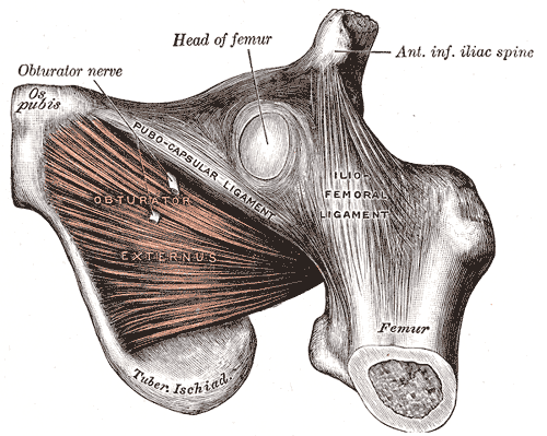
 The Obturator externus is a flat, triangular muscle, which covers the outer surface of the anterior wall of the pelvis. It arises from the margin of bone immediately around the medial side of the obturator foramen, viz., from the rami of the pubis, and the inferior ramus of the ischium; it also arises from the medial two-thirds of the outer surface of the obturator membrane, and from the tendinous arch which completes the canal for the passage of the obturator vessels and nerves. The fibers springing from the pubic arch extend on to the inner surface of the bone, where they obtain a narrow origin between the margin of the foramen and the attachment of the obturator membrane. The fibers converge and pass backward, lateralward, and upward, and end in a tendon which runs across the back of the neck of the femur and lower part of the capsule of the hipjoint and is inserted into the trochanteric fossa of the femur. The obturator vessels lie between the muscle and the obturator membrane; the anterior branch of the obturator nerve reaches the thigh by passing in front of the muscle, and the posterior branch by piercing it.
The Gemelli are two small muscular fasciculi, accessories to the tendon of the Obturator internus which is received into a groove between them.
The Gemellus superior, the smaller of the two, arises from the outer surface of the spine of the ischium, blends with the upper part of the tendon of the Obturator internus, and is inserted with it into the medial surface of the greater trochanter. It is sometimes wanting.
The Gemellus inferior arises from the upper part of the tuberosity of the ischium, immediately below the groove for the Obturator internus tendon. It blends with the lower part of the tendon of the Obturator internus, and is inserted with it it into the medial surface of the greater trochanter. Rarely absent.
The Quadratus femoris is a flat, quadrilateral muscle, between the Gemellus inferior and the upper margin of the Adductor magnus; it is separated from the latter by the terminal branches of the medial femoral circumflex vessels. It arises from the upper part of the external border of the tuberosity of the ischium, and is inserted into the upper part of the linea quadrata—that is, the line which extends vertically downward from the intertrochanteric crest. A bursa is often found between the front of this muscle and the lesser trochanter. Sometimes absent.
Nerves.—The Gluteus maximus is supplied by the fifth lumbar and first and second sacra nerves through the inferior gluteal nerve; the Gluteus medius and minimus and the Tensor fasciæ latæ by the fourth and fifth lumbar and first sacral nerves through the superior gluteal; the Piriformis is supplied by the first and second sacral nerves; the Gemellus inferior and Quadratus femoris by the last lumbar and first sacral nerves; the Gemellus superior and Obturator internus by the first, second, and third sacral nerves, and the Obturator externus by the third and fourth lumbar nerves through the obturator.
Actions.—When the Glutæus maximus takes its fixed point from the pelvis, it extends the femur and brings the bent thigh into a line with the body. Taking its fixed point from below, it acts upon the pelvis, supporting it and the trunk upon the head of the femur; this is especially obvious in standing on one leg. Its most powerful action is to cause the body to regain the erect position after stooping, by drawing the pelvis backward, being assisted in this action by the Biceps femoris, Semitendinosus, and Semimembranosus. The Glutæus maximus is a tensor of the fascia lata, and by its connection with the iliotibial band steadies the femur on the articular surfaces of the tibia during standing, when the Extensor muscles are relaxed. The lower part of the muscle also acts as an adductor and external rotator of the limb. The Glutæi medius and minimus abduct the thigh, when the limb is extended, and are principally called into action in supporting the body on one limb, in conjunction with the Tensor fasciæ latæ. Their anterior fibers, by drawing the greater trochanter forward, rotate the thigh inward, in which action they are also assisted by the Tensor fasciæ latæ. The Tensor fasciæ latæ is a tensor of the fascia lata; continuing its action, the oblique direction of its fibers enables it to abduct the thigh and to rotate it inward. In the erect posture, acting from below, it will serve to steady the pelvis upon the head of the femur; and by means of the iliotibial band it steadies the condyles of the femur on the articular surfaces of the tibia, and assists the Glutæus maximus in supporting the knee in the extended position. The remaining muscles are powerful external rotators of the thigh. In the sitting posture, when the thigh is flexed upon the pelvis, their action as rotators ceases, and they become abductors, with the exception of the Obturator externus, which still rotates the femur externally.
Media Attributions
- image434 © Henry Gray is licensed under a Public Domain license
- image436 © Henry Gray is licensed under a Public Domain license

