Foot & Ankle
19 Foot/Ankle Arthrology
Adapted from Henry Gray (1821–1865). Anatomy of the Human Body. 1918.
Talocrural Articulation or Ankle-joint
Lateral ankle ligaments
|
The ankle-joint is a ginglymus, or hinge-joint. The joint is formed by the lower end of the tibia and its (medial) malleolus, the (lateral) malleolus of the fibula, and the transverse tibiofibular ligament, which together form a concave mortise that receives the upper convex surface of the talus and its medial and lateral facets. The bones are connected by the following ligaments:
|
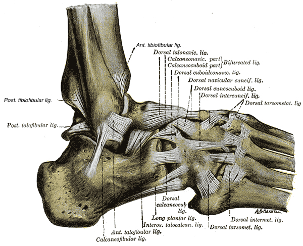
Anterior Talofibular Ligament.—The anterior talofibular ligament, the shortest of the three lateral ligaments, passes from the anterior margin of the fibular malleolus, forward and medially, to the talus, in front of its lateral articular facet.
Posterior Talofibular Ligament.—The posterior talofibular ligament, the strongest and most deeply seated, runs almost horizontally from the depression at the medial and back part of the fibular malleolus to a prominent tubercle on the posterior surface of the talus immediately lateral to the groove for the tendon of the Flexor hallucis longus.
Calcaneofibular Ligament. —The calcaneofibular ligament, the longest of the three, is a narrow, rounded cord-like ligament, running from the apex of the fibular malleolus downward and slightly backward to a tubercle on the lateral surface of the calcaneus. It is covered by the tendons of the Peronæi longus and brevis.
The Articular Capsule (capsula articularis; capsular ligament).—The articular capsule surrounds the joints, and is attached, above, to the borders of the articular surfaces of the tibia and malleoli; and below, to the talus around its upper articular surface. The anterior part of the capsule (anterior ligament) is a broad, thin, membranous layer, attached, above, to the anterior margin of the lower end of the tibia; below, to the talus, in front of its superior articular surface. It is in relation, in front, with the Extensor tendons of the toes, the tendons of the Tibialis anterior and Peronæus tertius, and the anterior tibial vessels and deep peroneal nerve. The posterior part of the capsule (posterior ligament) is very thin, and consists principally of transverse fibers. It is attached, above, to the margin of the articular surface of the tibia, blending with the transverse ligament; below, to the talus behind its superior articular facet. Laterally, it is somewhat thickened, and is attached to the hollow on the medial surface of the lateral malleolus.
| Deltoid Ligament (ligamentum deltoideum; internal lateral ligament) (Fig. 331).—The deltoid ligament is a strong, flat, triangular band, attached, above, to the apex and anterior and posterior borders of the medial malleolus. It consists of two sets of fibers, superficial and deep. Of the superficial fibers the most anterior (tibionavicular) pass forward to be inserted into the tuberosity of the navicular bone, and immediately behind this they blend with the medial margin of the plantar calcaneonavicular ligament; |
Medial Ligaments
|

The middle (calcaneotibial) descend almost perpendicularly to be inserted into the whole length of the sustentaculum tali of the calcaneus; the posterior fibers (posterior talotibial) pass backward and lateralward to be attached to the inner side of the talus, and to the prominent tubercle on its posterior surface, medial to the groove for the tendon of the Flexor hallucis longus. The deep fibers (anterior talotibial) are attached, above, to the tip of the medial malleolus, and, below, to the medial surface of the talus. The deltoid ligament is covered by the tendons of the Tibialis posterior and Flexor digitorum longus.
Midfoot ligaments, plantar surface

Gray’s Anatomy, Henry Gray, Illustrator: Henry Vandyke Carter
Media Attributions
- lateral ankle lig henry gray edited with tibiofib © Henry Gray
- image354 © Henry Gray is licensed under a Public Domain license
- Gray358 © Henry Gray is licensed under a Public Domain license
